Josef Trapani: Thank you all for coming. So we have a beautiful day for commencement weekend and I'm delighted and honored to get to introduce David P. Corey Back to Amherst campus. He's a graduate of the Amherst class of 1974. Bertarelli Professor of Translational Medical Science in the Department of Neurobiology at Harvard Medical School. Professor Corey is a dedicated investigator of the mechanics of hearing. Over decades of path-breaking work, the Corey Lab has demystified auditory transduction, the mechanism by which the delicate hair cells in our inner ear convert sounds and movements into electrical signals. In doing so, Professor Corey and his collaborators have transformed our understanding of hearing and laid the groundwork for precision targeted therapies to treat hearing loss. I believe we will hear more of this work today. The impact of Professor Corey's research is hard to overstate. Hearing loss is the most common form of sensory impairment in humans affecting more than 460 million people worldwide.
Josef Trapani: Hair cells, like car engines, are complex machines that need to be studied as they are running. Professor Corey has said, you can't figure out how a piston or a spark plug works by itself. You have to modify the part, put it back in the engine, and then gauge its effect on performance. An attraction to tackling complex problems with many moving parts has been evident since his days as an undergraduate physics major here at Amherst, when, for his senior thesis in Professor Gordon's lab, who's is up in the front row, he built an audio frequency maser using the nuclear Zeeman transition and helium three. Author of more than 150 scientific papers, Professor Corey has been widely recognized for his research, teaching and mentorship. Concurrent with his long career at Harvard Medical School, he has worked as a physiologist and neurobiologists at Massachusetts General Hospital and as an investigator at the Howard Hughes Medical Institute.
Josef Trapani: Prior to these appointments, he was a postdoctoral fellow and assistant professor of molecular neurobiology at the Yale School of Medicine in addition to a BA in physics from here at Amherst. He earned a Ph.d. In neurobiology from the California Institute of Technology. Even though he was a phyics major here at Amherst, he's played a big role in current neuroscience majors. This is the neuroscience textbook for the neuroscience majors in their intro course. And they have to read this chapter closely cause I also work in hearing and auditory and hair cells. And so we learn a lot about that portion of neuroscience, and one whole page is dedicated to Professor Corey including a photo shot right there of them. So they all see this as they go through the neuroscience major. So despite being a physics major, while here we think of you as an honorary neuroscience major and we actually have at Amherst College neuroscience sweatshirt for you. [laughter] That's great!
Josef Trapani: That's great!
Josef Trapani: And that's the mammoth with a brain on it!
David Corey: That's wonderful. Thank you.
Josef Trapani: Please join me in welcoming Professor David P. Corey, who will tell us more about how we hear, how we lose hearing and what we can do about it.
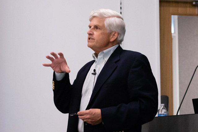
David Corey: All right, Joe, thank you very much for that kind introduction. I see a lot of about to be graduates in the audience and I know that some of you are going off to jobs and some are going off to graduate school. And for many of you that path seems clear and straight ahead. And as Joe mentioned, I started in physics and I'm going to end up talking to you about gene therapy. And so it's a little bit, maybe a little bit instructive to see how I really didn't take that straight a path after Emerson and maybe a little bit instructive to tell you exactly how I came to work on what I'm working on. So I loved physics at Amherst. I had wonderful professors such as Professor Cordon. I particularly liked electronics and instrumentation, but I realized by my senior year that it wasn't really quite for me, that I probably wasn't going to be that great a physicist and started looking around for other fields where I might be able to use that.
David Corey: And it was my great, good luck that Steve George came the fall of my senior year to start a new program in neuroscience. He was the first professor in what would eventually become a major. And he basically drew a number of disciples from students of different departments and we had discussion groups and meetings. And this started to look pretty interesting. But by the middle of my senior year, it was too late to apply to graduate school. I wouldn't have gotten in any way because I didn't have the preparation. And so Steve suggested that I might work for a year in a neuroscience lab, see if I like it. And then I might go on to graduate school. So when I went home for spring vacation senior year, Steve gave me a list of neuroscientists in the Boston area and I should look them up and see if I could work with them.
David Corey: Here's one of them. This guy, I just phoned him up. This is Torsten Wiesel. I'd never heard of him. He was then the chair of the neurobiology department at Harvard Medical School and would go on with David Hubel to win the Nobel Prize for his work in understanding how the brain processes visual information, and Torsten talked to me for half an hour and said, let me see what I can do and walked down the hall and asked Ann Stuart, a young assistant professor in the department, whether she needed a technician and she did, and suddenly I had a job. So, I needed to finish up. I went back to Amherst. As Joe mentioned, this was my senior thesis. It didn't actually work. We never quite achieved the sensitivity that we needed, but I did learn a huge amount in doing the project and that at least was enough to finish up the physics degree.
David Corey: And I had my diploma. And then went off to Harvard Medical School. So at Harvard Medical School in Ann's lab, we worked on this organism. This is the giant barnacles Balanus nubilus. And we worked on the visual system of the barnacle, and I will venture to guess that even the biologist among you don't know that barnacles have eyes. But they do, three of them. And it turns out very to be very useful to have an eye if you're a barnacle because if a fish passes overhead and casts a shadow, it's really good to bring in your filter feeding [inaudible ]before the fish nibbles it off. And they also have a very interesting way in which the light-sensitive cells, the photoreceptors, connect to at least what there is of a barnacle brain. And that was what we were studying. In any case, the neurobiology department at Harvard, which was the first department of its kind in the country, was a fantastic place at that time, probably the most exciting neuroscience collection of neuroscience researchers in the world.
David Corey: And I just loved it. So the whole field was interesting. The people were great and it became clear that I should try to go to graduate school. There was just one trouble. I had never studied biology at Amherst and not even in high school. And nevertheless, the number of graduate schools actually admitted me with no background. And I was finally admitted to Cal Tech. And the thing was that I was kind of sad and leaving physics because I love the electronics. I love the methods of studying physics. And I thought I would be leaving that all behind to go into neuroscience. And as you can see, this would not be the first time that I was profoundly mistaken. As it happened, the cells that we worked on these are the cells from the inner ear and their job is to convert a mechanical movement into an electrical signal.
David Corey: They do the first fundamental transformation from an acoustic wave into something the nervous system can understand, but they're phenomenally sensitive and fast. So this is about eight micrometers from top to bottom. This is actually from the inner ear of a bullfrog, but this cell, there's the top of one cell, can easily detect movements of a few billions of a meter, even as little as a few tenths of a billionth of a meter. And they can do to the detection on a timescale of microseconds and understand all of that. We had to use all the tools of physics. We had to make really fast pH electric stimulators that could move these cells and in a few tenths of a millisecond, use capacitance, compensated electronics in order to adjust for the electrical properties of the tissue in order to record the speed. And we're actually able to learn a lot using the tools that I had been able to bring from my Amherst background.
David Corey: So are there any lessons in this? I think one is that, that you shouldn't underestimate the role that luck will play in a career. I mean, if Steve George had come just one year later, my life would've been completely different. Another, I suppose, is to recognize when luck presents herself and be able to take advantage of that. And also kind of together with that, be willing to jump into fields where you're completely unqualified. But the corollary though is that you have to become qualified very quickly. And that means that you can never stop learning. And you have to spend your whole life learning, which of course is what Amherst prepared you for. So let me now tell you a little bit about what we learned and what we've learned over the last four decades. And in each case, what we'll be doing is asking the very same question of these cells, but going into a finer and finer level on a distant scale that smaller and smaller moving about one order of magnitude per decade.
David Corey: So this is the way the ear works and I'll start sort of at the large scale and work down. Here's a picture of my head. If you cut it about that way, and you can see, here's the pin of it, the external ear that collects sound. Many of you know about this, the sound is channeled through the ear canal where it makes the eardrum, the tympanic membrane vibrates back and forth. This vibration is coupled through the three bones of the middle ear, the auditory ossicles until finally this last bone called the stapes vibrating back and forth creates pressure fluctuations inside the inner ear. Now the inner ear actually contains two separate organs. So we have one, this snail-shaped cochlea. Now we're at a distance scale of a few millimeters and the cochlea subserves our sense of hearing. There are also these organs, the three semicircular canals and then two organs called the saccule and utricle and they use the very same type of receptor cell, but they subserve our sense of balance and very often than things that affect our hearing affect our balance just as well.
David Corey: Within these two organs are really a fairly small number of cells in the cochlea. The receptor cells that do this conversion occur in kind of a narrow ribbon that spirals along the center of each term of the cochlea, perhaps 16,000 cells in each year, which is very few compared to the hundred million photoreceptors that you have in each eye. And then similarly there are patches of receptor cells in these three or five organs of balance. Finally, they're also receptor cells in aquatic animals positioned along the wall of the body and those are the hair cells that Professor Trapani studies. So let's go a little bit further in what kind of cross-section through the cochlea, right about there. And then in this one slice of cochlea, you can see that there are these fluid-filled spaces. And then these receptor cells that we're interested in are just those four in each section.
David Corey: You can see one here and then three or four here. So those are the cells that we care about, but there's another cell type in here that we also care about. And those are the nerve cells that carry the information from the hair cell to the brain. The hair cells don'... these are the receptor cell--don't go to the brain directly, but their information is shuttled by what are called spiral ganglion neurons. And then that goes eventually to the auditory part of the brain stem and then to the rest of the brain. So if we zoom in a little bit farther, you can see again what are called these cells that are called hair cells. And I'll explain in a moment, you see one that's closer to the inside. And so that's called an inner hair cell. And a single row of those.
David Corey: This is what the top of an inner hair cell looks like. We're now down at a distance scale of about 1000000th of a meter, one micron. You can see that these are two or three micrometers tall and they're arranged in almost like a row of pipe organs here. The other group of cells, these outer hair cells have a fairly similar arrangement of these protrusions coming from the top. Again, there are about a hundred of these on the top of each cell and these are called stereocilia, not because they're like the true cilia that you know of in the lung or oviduct or in any place where a beating psyllium might move things along. Instead, these are full of actin filaments, but they're called cilia just because they look a little bit like the motile cilia that were better know. So we'll be interested in how these stereocilia might be part of a mechanism for the cell to detect a vibration.
David Corey: So it had also been known for a long time that as the sound is brought in through these middle ear bones to create a fluctuation in pressure in the cochlea, that each time the stapes moves in, it increases the pressure in this compartment and these hair cells are located on a fairly flexible membrane here so that with every positive phase of the pressure wave, this is pushed down and with all the hair cells riding on it and in the very next half cycle with a little a negative pressure, then this moves up so the whole apparatus is vibrating up and down at a hundred or a thousand times a second in your own ears as you listened to me. Now there's another part of this which is called the tectorial membrane. You can see this structure here that's actually made by cells over on this side and this tectorial membrane kind of sweeps over the region of the hair cells and it contacts the very tips of the stereocilia.
David Corey: And so now you can see as if this is moving up and down. Then every time this moves up, just because of the geometry of the system, the stereocilia will be bent one way and then the other and back and forth and each time bent forward and backward with each cycle of sound. So somehow, and again, the central question: How is it that the movement of the stereocilia could cause an electrical change inside the hair cell that the nerve fibers within carried to the central nervous system? Well, we know a little bit about how electrical changes are generated in nerve cells and other kinds of cells. And in general, this change is mediated by a type of protein that's called an ion channel. So this is a protein that fits in the cell membrane and it has a kind of a pore through the middle of it. It Can connect the outside of the cell to the inside of the cell.
David Corey: And the trick is, as many of you know, is that this pore tends to be fairly narrow and tends to select only some ions so that if the pore opens up, then charged atoms like sodium and potassium might flow into the cell. And that would change the voltage inside the cell. So first question we really need to know is how is it that the movement of these stereocilia would open and close an ion channel? The first clue came from asking simply how quickly do the channels open. And so we developed instrumentation where we could take away the tectorial membrane, touch the tips of the stereocilia themselves, and then in about a 10th of a millisecond we could move the stereocilia over, hold them, and then maybe a millisecond and a half later bring them back. And we can simultaneously measure the electric current that's flowing through a group of hair cells.
David Corey: And you can see here that the electrical current turns on very quickly after the movement of the bundle. If you were to fit a line to that and sort of characterize the opening speed, you would see that for small deflections of the bundle, it opens in about 500 microseconds in about a half a thousandth of a second. But if you give a larger deflection, it opens even faster and about a hundred microseconds. And now there's a second thing that you need to know, which is first of all that these experiments were done in a bullfrog, an animal not exactly known for speed, and the other thing is that to do the measurements we had to cool the tissue down almost to freezing to slow the whole thing down so that we'd had a chance of measuring it. And if you warm it up to a mammalian temperature to the temperature that your hair cells are working at right now, the whole thing is about 10 times faster.
David Corey: So it can open and close on a timescale of 10 or 20 microseconds, 20 millions of a second. This is great if you're a bat or a dolphin that wants to hear to 70 or even 100-kilo hertz in a dolphin ear, these stereocilia are moving over and back every 10 microseconds, you know, thousands of times a second. And that's great. So these ion channels, wherever they are, can open and close fast enough for the animal to be able to hear really high frequencies. But for understanding how the cell works, it had a completely different meaning. And many of you know that there are systems in biology where you might have one protein that contacts another one that might enzymatically modify a substrate, and then that activates something else. And in general, these kinds of enzymatic steps are slow. They might take a 10th of a second or a hundredth of a second, but none of them work as fast as a hundred thousandth of a second.
David Corey: And so what that was telling us was that none of these kinds of chemical systems could be involved in opening the ion channel instead. There had to be a very direct, probably mechanical connection between the movement of the cilia and the opening of the ion channel. You might almost think of something pulling directly on the ion channel. A second clue to this basic understanding came from the chips of bundles of stereocilia in different species. So here's one from a mammalian cochlea. Here's one from those hair cells on the side of a fish. The stereocilia are just these little guys right here. Here's one from the inner ear of a bullfrog. Here's one from the balance system of a mouse. And what you can see, even though they all have different shapes, is that in every case, the stereocilia are arranged in a series of graded heights from short to kind of medium to tall, short, taller, taller, taller, short and so forth.
David Corey: And so that must be something fundamentally important about the way the cell works, that that never changes and all vertebrate hair cells, experiments that were all that were done, not by me but by others in the lab at Caltech showed that these kind of hypothetical ion channels that were opened by movement at the bundle. We're actually located in the very tips of the stereocilia, not along the side, not at the bottom. And so we kind of had to figure what is it at the tips that might be special that might give us a clue. And an Australian anatomist, Jim Pickles, looked very carefully at the tips of the stereocilia and he noticed that there were these very fine filaments connecting the tip of one stereo psyllium to the side of the next taller one. Here's another one. There's one maybe here. One may be here and he called these tip lengths.
David Corey: Not a terribly imaginative name, but he was, he was clever and he knew what that might mean for the function of the cell. We can look at a tip link. This is a scanning electron micrograph that looks at the surface of the cell and to look a little bit better, we can make a slice through the cell and use a transmission electron micrograph to kind of see the inside. Here's the top of one cell and the side of another cell. You can see these actin filaments that make the stereocilia very stiff and there's the tip link just right along there. Not much more than a 10th of a millionth of a meter in size. And we know now that this has made of just a couple of protein molecules. So Pickles knew that the channels were at the tips of the stereocilia and he discovered these links at the tips of the stereocilia and he knew also how a bundle of stereocilia move when stimulated either by sound or by the experimenter.
David Corey: And you can see here we've taken the cells out of the animal, put them in a dish, put a probe on to the bundle and moved it back and forth. And you can clearly see that these cilia don't bend, but they pivoted their base and they stick together near their tips. So every time the bundle moves, this way, the tips are going to become a little bit further apart. And every time they move this way, the tips will come a little bit closer together. And so all you have to do is suppose, as Pickles, did that every time the bundle moves towards the tall stereocilia, the tip link is going to be stretched. And what he proposed then was this very simple model that the tip link might be directly connected to some sort of an ion channel. And every time you move this way, the tickling gets tighter, that pulls the channel open. And that is all there is in essence to the conversion of a vibration to mechanical stimulus. Every time the channel opens, you get a little pulse of current coming down through the stereocilia into the inside of the hair cell, changing the voltage inside the hair cell, and then allowing communication to the next cell online. So that's fairly satisfying, but it didn't really tell us what this channel was or really what any of the molecular components of this apparatus might be. So how could we figure that out? The answer in great part came from human genetics, which is tragic in a sense, but as geneticists began to identify families who had inherited deafness, where there might be a mutant copy of a gene that's passed on from parent to child, they would be able to figure out what the gene is and then ask what is the protein that the gene is the instructions for?
David Corey: And then ask where is that protein actually located? And for a great many of these different kinds of hereditary deafness. The protein was encoded by that chain, turned out to be near the tips of the stereocilia. So here a number of different proteins, if a mutation is in any one of them then the hearing apparatus doesn't work and the child is deaf. Let me point out a couple of them. This one's called cadherin 23. It forms the upper two-thirds of the tip link and a mutation in this gene causes profound congenital deafness. Kids are born completely deaf and their hair cells in the balance organ don't work either. There they have no intrinsic sense of which way is up and which way is down. So how do you learn to walk if you don't know which way's up and down and actually, you can do that.
David Corey: You can feel the floor on your feet, you can see the floor, you can see the ceiling. And then when they're about 20 years old, they started going blind as well and then are almost completely blind by the, by the middle of their thirties. Protocadherin 15 is another protein that's part of the tip link and that causes exactly the same symptoms if there's a mutation in that. So one by one we began to build up an idea of the different proteins that are part of this apparatus. It's a little bit, I suppose, like being given a box of watch parts and now your job is to try to put them all together to see how they might fit to form a functional machine. I want to concentrate on one that we've been particularly interested in. And this is the ion channel itself.
David Corey: I mean, after all, that is the core of how these cells would convert sound to a nerve signal. And that also was discovered by looking at hereditary deafness. In particular, there was a family in Southern India, and a couple of their children went to a school for the deaf in India, a boy and a girl. The parents had no deafness. And people at the school realize that these two kids came from the same family and they both had the same kind of deafness. And on further inquiry found that there was another child, another sister at home who was also deaf in the same way. And they started to ask whether this might run in families that turned out there was an uncle as well, the brother of the father, and he was deaf. And as they started to go back four or five generations, then two more kids turned up who were deaf.
David Corey: Now geneticists love this kind of a family tree because for every different individual in the tree, they can sequence the DNA, compare the DNA from individual to individual and then find maybe one letter of DNA that's different in the kids that have the deafness from all the ones that don't. Similar families were identified in other countries that seem, or the genes seem to be in the very same place in the genome. And it was eventually shown that this gene was one that they named TMC1 transmembrane channel-like, and we now have a lot of evidence that this TMC1 is actually the, the gene makes the directions for making this ion channel that is essential for hearing. This is the DNA directions for making TMC1 and many of you know that you can decode this. Every three letters of DNA indicates another amino acid and you can just read that out like you almost read the telephone book and figure out what the order of different amino acids is in the protein that the cell makes from these instructions. And I have a string of beads that are like amino acids and we can start reading the entire sequence one by one every three bases and figure out exactly what the amino acid sequence is for TMC1. And we just keep getting letters and letters and letters. Here is the complete arrangement of the letters. And at some point we noticed that some of the letters, that is, some of the amino acids are a little bit different from the others. Some of them we know are amino acids that love to live in membranes. And so we can kind of say, well maybe this is a part of the protein that is in the membrane of the cell and then maybe this is a loop outside.
David Corey: Or maybe this is a loop inside, but this doesn't tell us anything about how the protein would come together to actually act as an ion channel in order to let this specific amount of current go into the cell to change the electrical signal inside the cell. So how do you, how do you figure that out? One of the things that we know about a number of other ion channels is that they often occur as a collection, a little set of similar or identical proteins. So here for instance is very simple. Ion Channel, this little bit here has about a 120 amino acids that is, there are about 2,500 atoms in that one. But every time you see it in a cell, you see that it's arranged with three other ones that are identical to it and they're arranged together. This is looking from the outside of the cell to create a little tiny hole down the middle.
David Corey: And that little tiny hole tends to be a very favorable place for these charged items, these ions to go through. So this is very similar to an ion channel that is involved in the generation of electrical signals in neurons. Here's another one. You can see there's one, two, three, four, five similar subunits. This is an ion channel that's used in activating muscle. When a nerve fiber comes down to a muscle, it sends a signal to this channel and then the muscle contracts. Here's another one. One, two, three, four, five, six different subunits. This is a channel that actually connects the inside of one cell to the inside of another cell. It's called the gap junction protein, but you can see a theme. Again, there's a pore through the middle that the ions can go through. So we thought if we can just figure out how many TMC molecules come together to make this poor, then we would have a sense of sort of how it works.
David Corey: And so we did a lot of biochemistry and use a series of different assays and asked in many different ways how many TMCs come together. And the answer was always two. We have two TMCs, just two that come together. So how, I mean, how do you take two of them and stick them together to make some kind of a central port? It really didn't make any sense. It was really perplexing. But it turned out that there are a few ion channels that have been, that are known, that also occur as a pair. There's this one here that was discovered by Chris Miller, and here's a sequence, that was identified by Rod Mackinnon and here's another one. So that's called CLC1. This is one called TMEM 16. And as we started to look at TMEM 16, it turned out that if you look very carefully, you can see that the arrangement of amino acids in TMEM 16 was fairly similar to the arrangement of amino acids in the TMC1 protein.
David Corey: That is to say they were distant cousins in evolution. But what we know about proteins that are descended from the same original protein is that they very often share a similar structure. Even if the similarity when you just read the letters is not that great. And luckily for us, the structure of this TMEM 16 protein had been solved actually several different times. So we thought we can just kind of line up the similar amino acids and TMC one with their partners in TMEM 16 and then from that figure out what the structure of TMC one might look like. And so we did that. The interesting thing that's really unusual about this protein is that the ions don't go through the middle where you might think that they come where two proteins come together. Instead, this copy of TMEM 16 and this copy of TMEM 16 each have their own pathway for ions to go through and it's not in the middle it's somewhere in this groove out near the edge of the protein.
David Corey: But so that was really strange. Either way, we could then guess about what the structure of TMC1 might look like. And here again, you can see each of these sort of spirally things is a group of amino acids that go from the outside of the cell to the inside of the cell. Those were the little green regions and this kind of model. And again, the idea would be that the ion current might flow through these two regions in a pathway that would be very similar to that of the TMEM 16. Well, that still had to be tested. I mean if this is really true if they had similar structures and if the ions in our channel actually went through this groove, then we ought to be able to test that. Specifically what we might be able to do is to change an amino acid in this pathway one at a time.
David Corey: And in each case, we would have to take the modified protein, put it back into the hair cell and then ask, does the current through the hair cell change at all when we changed the amino acids. And in every case where we changed an amino acid in what we thought was the pathway for ions, the flow of ions did actually change and we changed amino acids somewhere else in the protein. The flow of ions didn't change at all. So this seemed to be very good evidence that the structure of TMC1 was pretty similar to what we were guessing based on this other protein. There's still a really big question that we don't know. And you know, this is something that I have to know before I die or well before I retire. And this is really the question at the very heart of the whole thing, which is how does the pore open and close, how is it that the tip link attaches to this protein?
David Corey: How is it that tension on the protein shifts or bands or unfolds or twists the protein to basically open and close that groove? And I just don't know. This is the thing that I have to figure out. I have a guess, but let me show you a further test that we did. And that is, and this was done by Marco Sotomayor, a colleague at Ohio state, which is to say, if the ions are really going through this groove, we ought to be able to model it mathematically, specifically, physically. And the way that you do this is to take all the atoms of the TMC molecule, about 14,000, put them into the computer, take a whole lot of atoms for the lipid molecules that make up the membrane, maybe another 30 or 40,000, all the water molecules. And then in red, a lot of potassium atoms that might be around there.
David Corey: And then you ask for every atom, what is the force that it exerts on its neighbors? And then you say, in the next 10th of a trillionth of a second, where is that out? I'm going to move. And you do this over again, over and over again. Every time calculating a 10th of a trillionth of a second. And then over time for hundreds of thousands of atoms, you can see how this protein might move and whether a potassium could actually go through it. So here is this movie and you can see now the little potassium ions running around and every once in a while one finds the opening zips down into the inside of the cell and another and another. And this is a process that would take place about 200 million times a second, slowly bringing enough electrical charge into the cell to change the voltage on the inside.
David Corey: And so this also gives us a lot of confidence that we understand how this problem works. So how would this actually come together? As I said, I don't know, but I could kind of, I'm going to guess that somehow the protocol here in 15 molecules come together as a pair that each one of them somehow hooks on to the TMC protein. And somehow by pulling here, it helps pull a section on that that will pull the channel open. But as I said, I just don't know.
David Corey: One of the wonderful things about being married to a scientist is that dinner time conversation is often about what we're studying in the lab. And my wife, who's here today is the human geneticist and a pioneer in gene therapy. And after 30 years of listening to this, she said, you know, when are you going to do something about this? When are you going to use this knowledge to actually help people that have a hereditary disorder? And so together we started to work on some strategies like that and I need to tell you first the different ways in which your ear might malfunction in order to understand how we might think about treating it. So here is one way that you can lose hearing. And this is sometimes called age-related hearing loss. Probably a third of people over 65 have some degree of hearing loss.
David Corey: And for some, it's really debilitating. And so that means there are about a million people a year in the United States that are entering the age when they're beginning to get some hearing loss. And of course, this often gets worse with age. What is the cause of it? In many cases it's noise trauma, it's exposure to rock concerts or power tools, noisy subways and things like that. And we can ask what happens to the inner ear if you play a loud noise to it. Here is again a scanning electron micrograph of an animal in which, it's just been in a quiet environment and you can see the beautiful arrangement of inner hair cells, three rows of outer hair cells, and it's a very, it's a pretty healthy ear. And then if you take that same animal and expose it to noise as loud as a chainsaw for just two hours, this is what the hair bundles look like.
David Corey: I mean, if they've just been trashed, it looks like something after Mount Saint Helens blew up to some extent the hair cell can repair that. But repeated insults of this sort eventually kill hair cells. There are other sources of other ways that you can kill hair cells. It turns out that there's certain drugs that will kill hair cells. Notable among them are some really powerful antibiotics that are part of what's called the immune glycoside family. And you've heard of them. Gentamicin, neomycin, kanamycin and those drugs will actually accumulate inside hair cells and because they can get in, they kill the hair cell. Cisplatin, a chemotherapeutic agent is also extremely ototoxic, meaning that it kills carousels. And so as we get older, what we eventually end up with is just many fewer hair cells. What's important to know is that hair cells are what we would call post-mitotic, meaning you have only the hair cells you were born with and probably not all of those and you never get any other ones.
David Corey: So this is one sort of hearing loss. It's extremely common. Another is more subtle and this is something that was worked out by my colleagues at Massachusetts eye and ear infirmary and they recognize that there were forms of hearing loss that didn't result from the death of hair cells, but instead, it was a more subtle disconnection of hair cells from the rest of the brain. And what we know is, is every time the stereocilia moved back and forth, we saw that there was an electrical charge inside the hair cell. The hair cell needs to send the signal to the nervous system. And so it releases a chemical called glutamate, a neurotransmitter that is received by the neurons and then activates those. But if you're dumping a lot of glutamate out in this region, the neuron say enough that I can't take it anymore. And they kind of retract their processes.
David Corey: So the hair cells are still there, the neurons are still there, but they're not talking to each other anymore. And this is been dubbed hidden hearing loss because if you ask somebody, can you hear this tone of a certain level? They, yeah, sure I can hear that. And it's because a few of the connections are still there. But if you ask, can you understand speech in say, a noisy room, that's much, much harder because maybe they've lost 80% of the information channels that are going to the brain. So that's another kind of hearing loss that is equally prevalent and only recently recognized. The third kind that I mentioned is really, I'm far less common but still devastating. And that is hereditary deafness. So it turns out that there are about 4,000 kids a year who are born with severe or profound hearing loss. And as many of you know, infants have to be tested before they leave the hospital to see if their hearing is normal so that there can be some therapy early on. If it turns out to be abnormal, in many cases, maybe the majority of cases, this is due to some sort of hereditary defect that is they carry a mutation in one or another gene that is in a critical, that in codes a critical protein for hearing. Now they're more than a hundred different genes that have been recognized who's protein products are critical for hearing. And one by one we began to figure out what the genes are, what protein they might make. This, although it's rare, is in some ways easier to treat than the other two. And that's what Sandra and I tried to do in developing a therapy. And so in the case of hereditary deafness, all the hair cells are defective. So what can be done about it?
David Corey: So this is my new friend. This is an adeno associated virus. It's really tiny. It's about 25 billions of a meter across. Another way of putting it is about 10 of these stacked side-by-side would be the diameter of a single stereo psyllium. And this is a favorite of people working in gene therapy because it's relatively nontoxic and in many cases it's pretty good at attaching to a cell, entering the cell. And then whatever DNA it has inside, it's released and that DNA goes into the nucleus and then the cell is trick. The cells think that that's its own DNA and then we'll make the protein that, that DNA and codes. Um, so this is sort of the way it's most simplistic. The way gene therapy might work, where the icosahedral structure of the adeno associated virus can be used to carry a particular gene.
David Corey: So, for instance, if you have two parents, each of them carrying the mutation, then a child might be unlucky enough to inherit two mutated copies. And in the case of a simple recessively inherited deafness then they would have a defective protein. So what we would do then is to take an AAV, put a normal copy of the gene into the AAV, deliver the AAV to the hair cells, and then see if that gene released from the AAV can now produce the normal protein in the cell, can function normally. Let me tell you about one example of this. And in this case, we're focusing on one of those different proteins that seems to be associated with the mechanotransduction channel. The TMC one itself, and this is a protein that shut, that has this awful name Lhf PL5, which doesn't really stand for anything, but I have at least learned to pronounce it.
David Corey: And the Lhf PL5 is kind of interesting as a protein. It's a small protein. It's also embedded in the membrane of the cell and it has four places where the protein threads back and forth across the membrane for the aficionados. There's an extracellular Beta sheet out here. And what we know is that mutations in any one of three places here, here or here in each case where a single amino acid will be changed to something inappropriate, any of those mutations would cause hearing loss. If you take a mouse and introduce a similar mutation, you can see that a normal mouse again has a beautiful arrangement of outer hair cells and inner hair cells. But a mouse carrying this mutation and Lhf PL5 has terribly disordered hair bundles and it's completely deaf. It also has essentially no sense of balance. So how can we use this kind of model system to see if we can develop a therapy that might later be used in humans?
David Corey: Well, all we really need to do is to make a virus, pack some of the right DNA into it and get it to the right cells so we can do a simple surgical operation, even on a newborn mouse and use a glass needle just to inject some virus into the cochlea. And it kind of accumulates in this space and diffuses up to the hair cells. And we found viruses that are now very good at delivering DNA specifically to her cells. The general strategy for the experiment has shown, here again, we take a bit of DNA, put it into the AAV, inject into a newborn mouse ear, and then we can test whether this has restored the function of the hair cells in a couple of different ways. In one of them we can wait five days so that the new protein is made and then take the cochlea out of the mouse and test it.
David Corey: And we actually have a really simple way of testing whether hair cells are working because this TMC one pore is big enough that if you put a small fluorescent protein, sorry, a small fluorescent molecule on the outside, it slips into the cell and makes every hair cell brightly fluorescent. So in a normal mouse, all the inner hair cells, not three rows of outer hair so are brightly fluorescent. In a mouse that has a mutation. In Lhf PL5 you really see no fluorescence. But if you take a similar mouse and inject the normal DNA into the year, now you're seeing that many, not most, but many at least of the hair cells. Now we'll let the dye in indicating that their ion channels are working properly.
David Corey: Another way that you can see if this is working is to test the electrical response near the brain of the mouse when sound is played to it. So you can basically play a little tone to a mouse and then put a little wire on the back of the head. It's sort of like a little micro EEG and every time the sound is played to the mouse, the signal goes from the cochlea to the brainstem and a tiny, tiny voltage, a few millionths of a volt are generated. But we can amplify those. And see whether the electrical response in the mouse as normal. So if you take a mouse that's perfectly normal and play a little Chirp, a little, maybe a two or three or five-millisecond tone, very, very quiet, just 10 decibels, doesn't really give you any electrical response. Make it 20 decibels, no. 30 decibels, just beginning to see a little bit of a wiggle. And as you go up each time, in order of magnitude in power, you see that this little electrical response gets larger and larger. So this mouse can hear perfectly well. In the mouse lacking lLhf PL5 no amount of sound will produce the electrical signal. The mouse is completely deaf. But if we take the mouse lacking Lhf PL5 and inject the proper DNA in, now you can see, well, you need to use a louder sound. But once again, you can get this electrical signal that is a kind of a fingerprint for normal hearing in the mouse.
David Corey: There's yet a third way to test the function of the ear. And remember that if you have a mutation that changes the hearing in the auditory part of the ear, it often changes the function of the balance part of the ear. But if we inject a virus in here to try and treat the hearing part, it can diffuse over to the balance part and we can ask if the balance is restored. How do you test balance in a mouse? A really simple way is to drop it into a tank of water and see if it can swim. Actually, it can swim fine. It's not very happy about it, but it knows which way is up, it knows which way is down. It has a sense of balance. And it can swim in order to keep its head above water. It can always get its head to the air. And it will swim. Its not very happy. And it'll go back and forth. But either way, this is a mouse that can swim perfectly well. I guess it's a little tired but it will keep going. If you take a mouse that is lacking in Lhf PL5, it has no way of knowing which way is up. And swims, goes every which way trying to find air and we scoop it out before anything happens. But now take a mouse that's been treated with this gene therapy vector, and it's not swimming quite as well but it's keeping its head above water. It's sense of balance has been restored.
Okay, we've done this now for three different kinds of hereditary deafness in a mouse model. In a couple of cases it a recessively inherited deafness where you have to get two copies of the gene. In another case it's a dominantly inherited deafness where instead of adding a normal copy of the gene, we use a kind of chemical scissors to cut the mutant copy of the gene, to recognize one nucleotide, one letter of the DNA that was incorrect out of the 3 billion in the mouse genome and snip that gene in order to get rid of the bad copy. Still, this is just very early days and there's the question of whether we can take this to humans anytime soon. And you can imagine gene therapies that might treat other forms of hearing loss. For instance so if you've lost a lot of hair cells, you might think about putting something into the ear that makes the cells start to divide again, and start to make new hair cells, and you might imagine restoring a full population of hair cells. In the case of the hidden hearing loss, you might think about putting something into the neurons that make that grow out or something into the inner hair calls that attract the neurons in order to restore the normal connections. But these are still a long way off.
So I think in terms of gene therapy, the future is really bright. We're still at a very early stage, but I'm very excited about what's going to be possible in the years ahead.
If I can just end with one little story, it really has nothing to do with science, but I thought you'd be interested in. To me, it's a wonderful honor to be here and to receive this award, and it happens that my father also went to Amherst College, the Class of ’41, he was editor of the Amherst Student. He went on to Harvard Business School, had a distinguished career and about 30 years ago, Amherst asked him to come for an honorary degree, but it happened that he had a vacation planned with my mother to go to Europe that week. The tickets had been bought. The reservations made, the deposit, and he loved Amherst and this was a great honor, but he loved my mother more, and so he had to decline the honor. So if I may, tomorrow I'll try to accept this on small part on my father's behalf as well.
[applause]